Event-related potentials provide better time resolution than do structural neuro-imaging. Although their principal disadvantage is poor spatial resolution, combining measurement of event-related potentials with structural neuro-imaging can provide valuable information on both temporal and spatial features of neurophysiological abnormalities in schizophrenia.
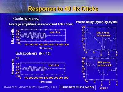
Dr. McCarley first described an event-related potential called gamma oscillation, measured by EEG recordings after presentation of a 40 cycle/second clicking stimulus. Measurement of gamma oscillation can detect abnormalities in the frequency at which brain regions communicate. Abnormalities of gamma synchronization in brains of schizophrenic patients may give rise to perceptual and cognitive abnormalities, such as aberrant semantic association, discontinuity in thinking, and/or hallucinations.
In normal subjects, the amplitude of the gamma oscillation response builds steadily and then fades after cessation of auditory stimulation. Schizophrenic patients show delayed onset of gamma synchrony followed by delayed termination of gamma synchrony after cessation of auditory stimulation. This is consistent with abnormal communication between brain areas.
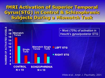
Another event-related potential is auditory mismatch, which is assessed by measuring brain electrical activity after tones of different frequencies. Comparison of responses to tones of different frequencies reflects ability to automatically process stimulus discrepancy. Subjects with schizophrenia have reduced mismatch negativity, particularly in the left hemisphere of the brain. This comports with a recent small study reporting a relationship between abnormalities of auditory mismatch and reduced activation of the superior temporal gyrus (on magnetic resonance imaging) in schizophrenic patients, compared with normal controls. These findings are consistent with auditory processing abnormalities in schizophrenia.
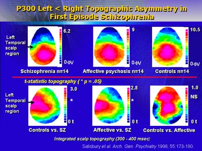
To measure another event-related potential called P300, subjects listen to typical and atypical tones and are asked to count the latter. The P300 is a positive electroencephalographic wave occurring 300 milliseconds after atypical tones. P300 in patients with a first psychotic episode reveals a deficit in the left temporal scalp region. This deficit is correlated with reduced gray matter volume in the posterior superior temporal gyrus. Normal subjects and subjects with mania show neither abnormality.
McCarley and colleagues plan to look next at P300 latency (interval between stimulus and P300 response), which may prove a useful marker for distinguishing auditory processing in normal and schizophrenic subjects.