| Continuous
administration of C-type natriuretic peptide (CNP) improved
left ventricular dysfunction and attenuated cardiac remodeling
after myocardial infarction. Mediators of these beneficial effects
may include direct inhibition of collagen synthesis and mural
hypertrophy, and acceleration of angiogenesis. These results
suggest CNP may be potentially useful as a cardioprotective
agent.
Left ventricular remodeling is a major cause
of heart failure and death after myocardial infarction. Dr.
Soeki and colleagues have previously shown that C-type natriuretic
peptide (CNP) inhibits DNA and collagen synthesis by fibroblast
more potently than atrial and brain natriuretic peptides in
vitro.
Because CNP appears to have potent inhibitory effects on
cardiac fibrosis and hypertrophy in vitro, investigators
hypothesized that CNP might attenuate the cardiac late remodeling
that can occur after myocardial infarction in vivo.
To assess this hypothesis, Dr. Soeki and colleagues observed
the effects of CNP on progression of cardiac remodeling in
a rat model of myocardial infarction.
The researchers used coronary artery ligation to induce experimental
myocardial infarction in male rats. Then, some of these rats
received a CNP infusion at 0.1 μg/kg per minute for 2 weeks,
beginning 4 days after the infarction. A second group of rats
received vehicle only, which consisted of a 5% glucose solution.
A third group of rats underwent a sham surgical procedure
with no coronary ligation.
To determine effects on systolic blood pressure and pulse
rate, investigators took measurements before myocardial infarction
and at 1 day, 1 week, and 2 weeks after infarction. As a result,
the systolic blood pressure and pulse rate were not perturbed
by CNP infusion at any time points. Investigators also performed
echocardiographic studies 4 and 18 days after the coronary
ligation or sham operation. They excluded rats from the study
that had fractional shortening of more than 20% or an E wave
to A wave ratio of less than 3 in the echocardiography performed
4 days after MI.
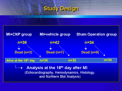
Final analysis on the 18th day of the study included 34 rats
that had an infarction and received CNP, 35 rats that had
an infarction but received only vehicle, and 34 rats that
underwent sham operation.
Although left ventricular end-diastolic dimension was greater
in the rats that underwent coronary ligation than in rats
that underwent the sham operation, CNP infusion did significantly
attenuate left ventricular enlargement.
The Effect of CNP on Echocardiographic Parameters
| |
Sham
|
MI+Vehicle |
MI+CNP
|
| AWT diastole, mm |
1.2±0.0
|
0.9±0.0**
|
0.9±0.0** |
| AW thickening, % |
71±1
|
10±1** |
10±1** |
| PWT diastole,
mm |
1.3±0.0
|
1.5±0.0**
|
1.4±0.0** ## |
| PW thickening, % |
70±2
|
42±1**
|
53±1** ## |
| LVDd, mm |
67±1
|
83±1**
|
77±1** ## |
| FS, % |
35±1
|
16±0**
|
18±0** ## |
| E velocity, cm/s |
88±2
|
112±3**
|
102±3** ## |
| A velocity, cm/s |
51±2
|
19±1**
|
26±1** ## |
| E/A |
1.8±0.0
|
6.2±0.2**
|
4.2±0.2** ## |
Values are mean±SEM.
**P<0.01 compared with the sham-operated
group;
##P<0.01 compared with the MI+vehicle group.
AWT=anterior wall thickness
AW=anterior wall
PWT= posterior wall thickness
PW=posterior wall
LVDd=left ventricular end-diastolic dimension
FS=fractional shortening
E=early filling wave
A=atrial filling wave |
|
They also found CNP treatment significantly improved systolic
and diastolic left ventricular dysfunction without affecting
arterial pressure. In the group that did not receive CNP,
left ventricular end-diastolic pressure was higher than in
the sham group, and the peak rate of contraction (dP/dtmax),
the peak rate of relaxation (dP/dtmin), and cardiac output
was lower.
The Effect of CNP on Hemodynamic Parameters
| |
Sham
|
MI+Vehicle
|
MI+CNP |
| HR, bpm |
412±
5 |
421±
6 |
410±
5 |
| MAP, mmHg |
120±
2 |
99±
2** |
103±
2** |
| LVSP, mmHg |
139±
2 |
116±
2** |
118±
2** |
| LVEDP, mmHg |
7±
0 |
18±
1** |
13±
1** ## |
| RVSP, mmHg |
38±
1 |
47±
1** |
45±
1** |
| RAP, mmHg |
2±
0 |
3±
0* |
3±
0* |
| LV dP/dt max, mmHg/s |
7970±
156 |
5019±
155** |
5743±
155** ## |
| LV dP/dt min, mmHg/s |
-6216±
158 |
-3791±
151** |
-4644±
147** ## |
| CO, mL/min |
98±2 |
73±
2** |
81±
2** ## |
| CI, mL/min/kg |
329±
6 |
278±
6** |
299±
6** ## |
Values are
mean±SEM.
**P<0.01, *P<0.05 compared with the sham-operated
group;
##P<0.01 compared with the MI+vehicle group.
HR = heart rate
MAP = mean arterial pressure
LVSP = left ventricular systolic pressure
LVEDP = left ventricular end-diastolic pressure
RVSP = right ventricular systolic pressure
RAP = right atrial pressure
LV dP/dtmax or min = peak rate of left ventricular
contraction or relaxation
CO = cardiac output
CI = cardiac index
|
|
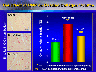
Dr. Soeki also reported that CNP attenuated increase of
morphometrical collagen volume fraction in the non-infarct
region.
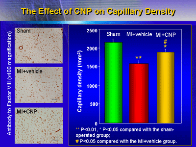
The CNP infusion also significantly improved capillary density,
which was reduced by myocardial infarction, in the non-infarct
region.
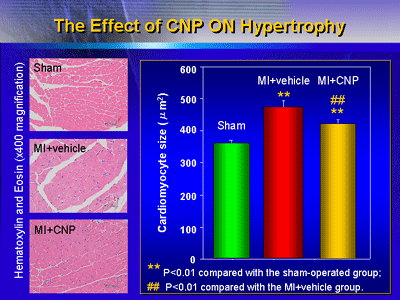
In addition, CNP prevented an increase in the heart weight
to body weight ratio and hypertrophy of the cardiomyocytes
in the non-infarct region.
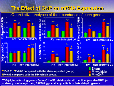
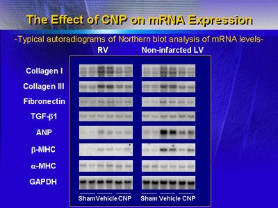
Finally, CNP suppressed increases of collagen 1 and collagen
3 mRNA levels associated with fibrosis in the non-infarct
region. The CNP infusion also suppressed increases of atrial
natriuretic peptide and β-myosin heavy chain mRNA levels associated
with cardiac hypertrophy.
These results suggest that in vivo CNP administration
can improve left ventricular dysfunction and attenuate cardiac
remodeling. The CNP infusion appeared to have beneficial effects
on cardiac performance. However, it had very little effect
on blood pressure, heart rate, and infarct size. This suggests
some mechanism other than hemodynamic improvement or reduction
in size of infarct would explain the beneficial effects of
CNP.
One possibility is that CNP directly inhibits myocardial
fibrosis. Another possibility is that CNP attenuates myocardial
hypertrophy after myocardial infarction. Some research suggests
CNP may activate the vascular endothelial cGMP/cGMP-dependent
protein kinase pathway; through this, CNP may improve the
reduced cardiac capillary density after myocardial infarction,
leading to less impairment of left ventricular function.
Regardless of the mechanism, the results of this study suggest
the potential usefulness of CNP as a new cardioprotective
agent.
|